Fig. 3.
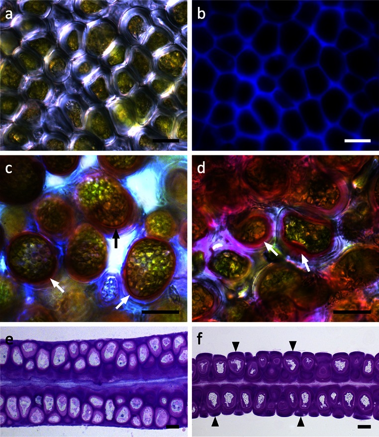
Light micrographs of U. compressa. a Surface view of a central area of the thallus. No intercellular spaces are visible between the polygonal cells. b Corresponding CFW staining of cellulose. c RR staining of fresh samples to visualize pectins, which are abundant in the inner cell wall layer attached to the protoplast (arrows). d RR staining in a sample desiccated for 30 min. The inner pectic-rich layer increases in diameter (arrows). e Chemically fixed, TB-stained semithin cross section of a fresh sample showing an irregular arrangement of cells in both thallus layers. TB-stained cross section of a sample desiccated for 30 min. Fragments of the cell wall matrix are attached to periclinal cell walls (arrowheads). The protoplasts show undulations followed by the cell walls, while no retraction of the cytoplasm from the cell walls occurs. The thickness of the thallus segment decreases. Scale bars 10 µm

