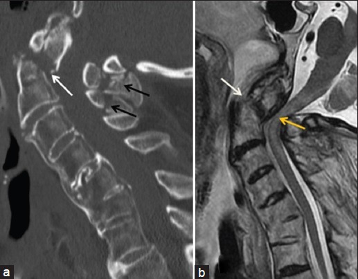
Figure 11.

Middle-aged adult with a history of ankylosing spondylitis and a Type II dens fracture with instability after trauma. (a) Sagittal reformatted CT image through the cervical spine reveals a displaced, comminuted fracture through the base of the dens. (white arrow), as well as comminuted posterior element fractures at C-2 and C-3 (black arrows). (b) Sagittal T2 MR image in the same patient demonstrates similar findings, along with severe central canal stenosis and cord compression with underlying cord signal abnormality (yellow arrow). There is also disruption of the anterior longitudinal ligament (white arrow).
