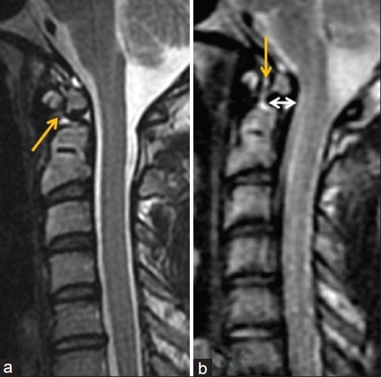
Figure 5.

28-year-old man with chronic neck pain and an os odontoideum with instability. (a) Sagittal T2 MR image through the cervical spine in the neutral position shows normal alignment of the os odontoideum with respect to the C-2 body and a normal atlanto-dental interval (yellow arrow). (b) Repeat imaging with passive flexion shows posterior displacement of the os odontoideum (white arrow) and widening of the atlanto-dental interval (yellow arrow).
