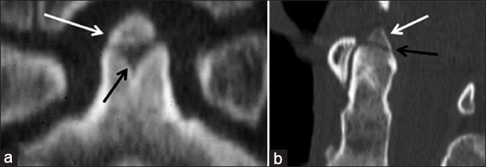
Figure 6.

10-year-old boy with a normal ossiculum terminale versus an elderly gentleman post-trauma with an acute Type I dens fracture. (a) Coned-down reformatted coronal CT image of the C-2 vertebral body in the young boy shows a smooth, corticated ossification along the superior margin of the dens (white arrow) with an underlying “V”-shaped cartilaginous cleft. (b) Sagittal reformatted CT image in the elderly trauma patient demonstrates an ossific fragment along the superior margin of the dens (white arrow) with a subjacent lucent fracture line (black arrow).
