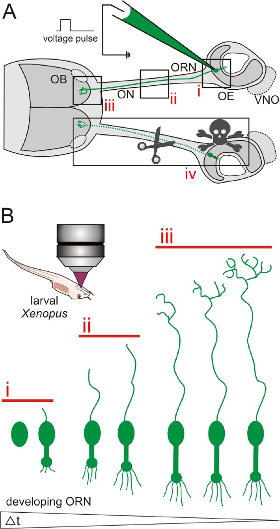
Figure 2.

Schematic illustration of the experimental advantages of the olfactory system of larval Xenopus laevis.
(A) Schematic representation of the peripheral olfactory system of larval Xenopus laevis. Olfactory receptor neurons in the olfactory epi-thelium (OE) of anesthetized larval Xenopus can be labeled via spatially restricted electroporation of e.g., fluorescent dyes. Stained cells can be visualized in the OE (i), their axons can be followed through the olfac-tory nerve (ON, ii), and their axon terminals identified in the olfactory bulb (OB) (iii). Introduction of lesions in the OE, the ON and/or the OB is easy (iv). The vomeronasal organ (VNO) and the accessory OB are outlined by dotted lines. (B) Labeled stem/progenitor cells or im-mature ORNs can easily be investigated in anesthetized larvae using a confocal or multiphoton microscope. Using in vivo time lapse imaging, early stages of neural stem/progenitor cell differentiation can be mon-itored in the OE (i), axon development can be tracked in the ON and the OB (ii and iii), and synapse formation can be observed in glomeruli of the OB (iii). Individual cells can be followed over long time spans (up to several weeks).
