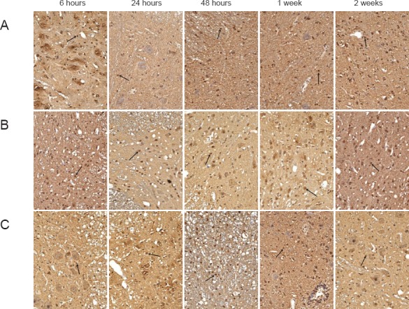
Figure 3.

Effects of hydrogen-rich saline on caspase-3 immunoreactivity in rats with spinal cord injury (immunohistochemical staining, × 200).
(A) SP group (sham-operated plus physiological saline); (B) SH group (spinal cord injury plus hydrogen-rich saline); (C) SSP group (spinal cord injury plus physiological saline). Nuclei of caspase-3-immunoreactive apoptotic cells (arrows) were stained brown, and positive cells could be seen at each stage in the SH and SSP groups. At 24 and 48 hours and 1 and 2 weeks after the injury, caspase-3 expression was greater in the SSP group than in the SH group.
