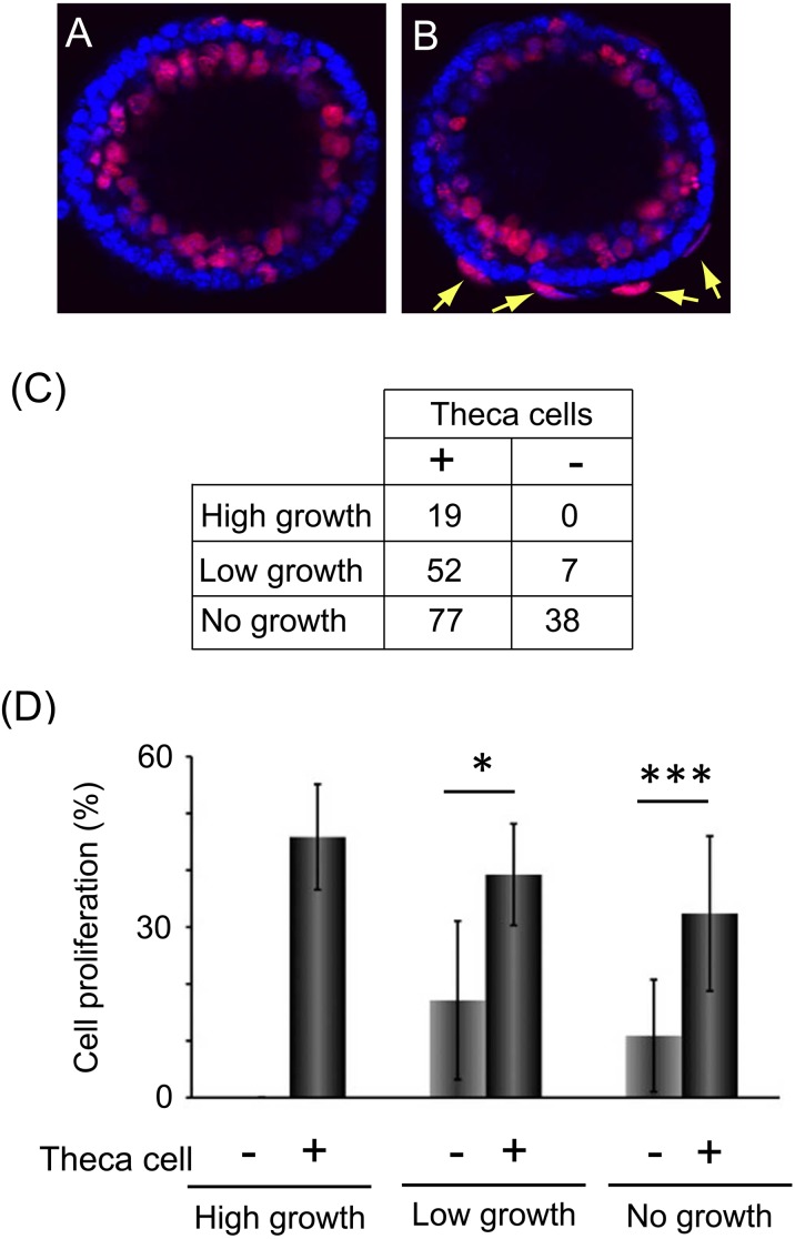
Fig. 3.
Theca cells promoted granulosa cell proliferation of secondary follicles. (A, B) The images show the granulosa cell proliferation around cultured secondary follicles stained using an EdU labelling kit (red) and Hoechst 33258 (blue). Panel A indicates follicles without theca cells, and panel B indicates follicles with theca cells (arrows). (C) The number of follicles containing theca cells (+) and no theca cells (–) in high-growth, low-growth and no-growth follicles collected from twelve mice. These follicles were used in subsequent experiments, the data for which are shown in Fig. 3D and Fig. 4A. (D) The graph indicates the rate of granulosa cell proliferation of each growing follicle. They were separated by whether they contained theca cells or not. Statistical significance is indicated by an asterisk (*P < 0.05, *** P < 0.001). Values are presented as means ± standard deviation. The numbers of samples used in this experiment were as follows: high-growth theca cell (–), 0; high-growth theca cell (+), 19; low-growth theca cell (–), 5; low growth theca cell (+), 29; no-growth theca cell (–), 19; no-growth theca cell (+), 51. These follicles were obtained from eight mice.

