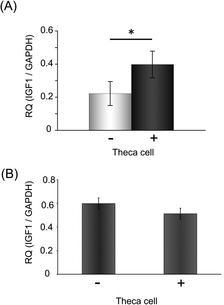
Fig. 5.
The theca cell layer increased the expression of Igf1 mRNA in the culture of secondary follicles in the no-growth group. (A) The expression level of Igf1 mRNA of cultured secondary follicles with theca cells was higher than in those without theca cells. The total mRNA of each group was extracted from ten follicles. (B) The expression level of Igf1 mRNA of culture granulosa cells. We compared the expression level of each group, cultured granulosa cells and co-cultured granulosa cells with theca cells. Statistical significance is indicated by an asterisk (*P < 0.05). Values are presented as means ± standard deviation.

