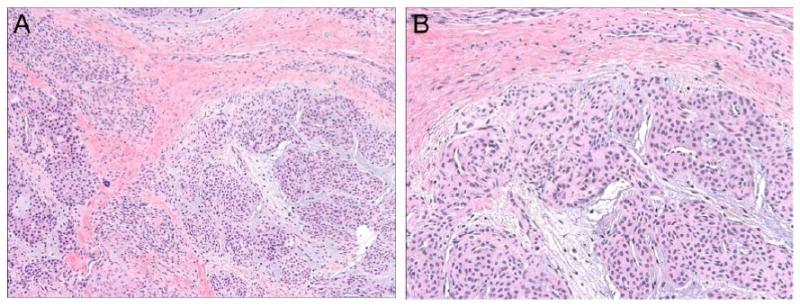
Figure 3. Typical histological features of glomus tumor.

Routine hematoxylin and eosin (H&E) staining demonstrates a perivascular, proliferation of homogenous round cells with round to ovoid nuclei arranged in multicellular layers around blood vessels. Lesional cells are set in a background of myxoid matrix with stellate cells. (A) 100 × magnification; (B) 200 × magnification.
