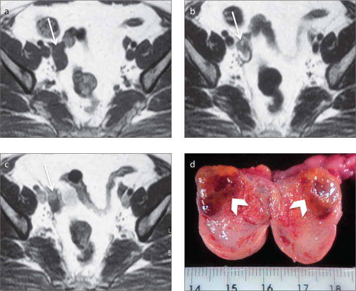
Figure 5.
a–d. A 53-year-old female patient, who was being monitored for hirsutism, diagnosed with a right ovarian Leydig cell tumor. Axial T1-weighted image (a) shows an increased in size right ovary (arrow). Axial T2-weighted image (b) demonstrates a hypointense, small solid lesion in the right ovary (arrow). Axial gadolinium-enhanced T1-weighted image (c) demonstrates enhancement of the lesion. Note that the hyperintense mass is well delineated against the ovarian stroma (arrow, c). The section surface of the right adnexal specimen (d) contains an ill-defined brown–yellow hilar tumor with a long axis of 10 mm (arrowheads).

