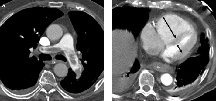
Figure 1.

a, b. A 82-year-old man presenting with hypoxemia, hypotension, sinus tachycardia and ECG-changes suggesting right ventricular strain. Multidetector CT (a) shows a saddle embolism, with signs of right ventricular dysfunction with high RV/LV diameter ratio of 2.8 (arrows in b; arrow in the LV cavity is partly overlying the papillary muscle). The patient was treated with thrombolysis which resolved the pulmonary embolism (b), however, the patient finally died of ventilation-associated pneumonia and septic shock.
