
Figure 3.
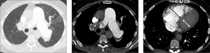
a–c. CTPA and HRCT reconstruction in CTEPH and mosaic perfusion. A 53-year-old man with CTEPH who presented with persistent dyspnea and dyspnea on exertion lasting several months, known with a history of adequately treated acute PE six years before. Blood analysis was unremarkable except elevated NT-proBNP (1212 ng/mL), echocardiography showed severe RV and right atrial dilatation with moderate tricuspid insufficiency and an estimated pulmonary artery pressure of 62 mmHg. Right-sided catheter measurements showed a pulmonary vascular resistance of 781 dyne·s/cm5 and a mean pulmonary artery pressure at rest of 46 mmHg. CTPA with high resolution reconstructions in lung setting (a) shows mosaic perfusion with hyperperfused pulmonary areas of high attenuation associated with larger vessels, and areas of hypoperfusion with low attenuation that contain smaller vessels. In soft tissue setting (b), the diameter ratio of the main pulmonary artery to the aorta is >1, indicative for pulmonary hypertension. Also note the wall-adherent thrombus and atherosclerotic calcification of the main pulmonary arteries, as signs of pulmonary hypertension. There is intraluminal web in the segmental artery to the left upper lobe. Dilated RV and flattening of the ventricular septum indicate RV dysfunction (c). Figure is published with patient’s permission and courtesy of Dr. I. Bahce and Dr. A. Boonstra, Department of Pulmonary Diseases at the VU University Medical Center Amsterdam, the Netherlands.