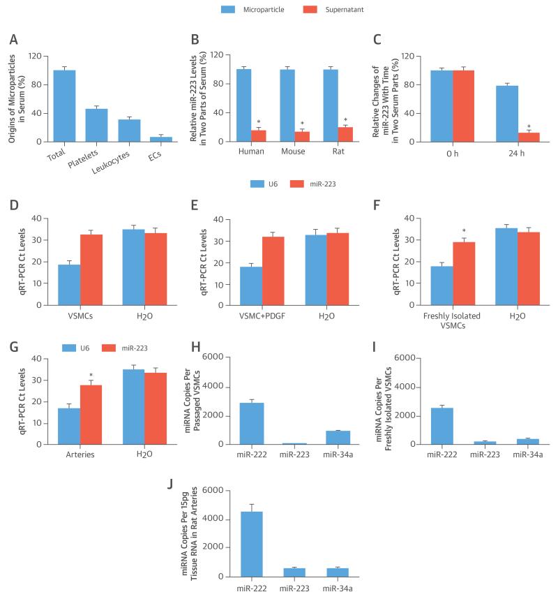
FIGURE 1. miR-223 in Circulating Serum, VSMCs, and Vessels.
(A) Microparticles arise from different sources in serum (n = 6). (B) The extracellular distribution of micro-ribonucleic acid (miR)-223 in 2 parts of serum. (C) MicroRNA-223 exhibits varying stability in microparticles and supernatant parts within 24 h. (D) The cycle threshold (Ct) levels of miR-223 and U6 in water (H2O) (negative control) and in passaged vascular smooth muscle cells (VSMCs) cultured in serum-free medium. (E) Platelet-derived growth factor (PDGF) (20 ng/ml) stimulation does not induce any miR-223 expression in passaged VSMCs. (F) A significant amount of miR-223 can be found in freshly isolated VSMCs. (G) A significant amount of miR-223 can be found in normal vascular walls. Absolute quantification of miR-223, miR-222, and miR-34a in (H) passaged VSMCs, (I) freshly isolated VSMCs, and (J) normal vascular walls. *p < 0.05 compared with control subjects. EC = endothelial cell.

