Figure 4.

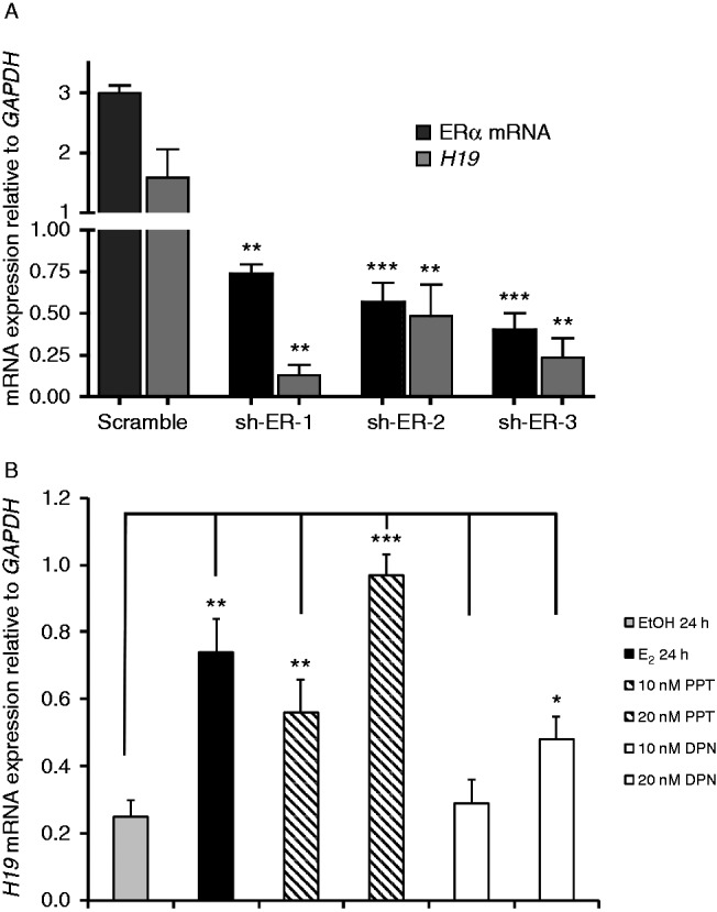
Estrogen-regulated expression of H19 is mainly regulated through ERα. (A) ERα transcript levels were decreased in MCF7 cells with three different sh-RNA fragments (sh-ER-1, -2, and -3) using lentiviral transduction. ERα and H19 transcript levels were examined in the transduced cells by qPCR. All of the data are normalized to the GAPDH transcript levels. (B) MCF7 cells were grown under estrogen-deprived growth conditions and treated with either EtOH, E2, PPT (a selective ERα agonist), or DPN (a selective ERβ agonist) for 24 h. H19 expression was ascertained using qPCR and was normalized with respect to GAPDH expression (n=3). Compared with EtOH, as little as 10 nm PPT increased H19 transcript levels, whereas DPN at the 10 nM concentration level had no effect. However, at 20 nM concentration, both PPT and DPN increased H19 expression (3.9- and 1.9-fold respectively). *P<0.05, **P<0.005, ***P<0.0005.

 This work is licensed under a