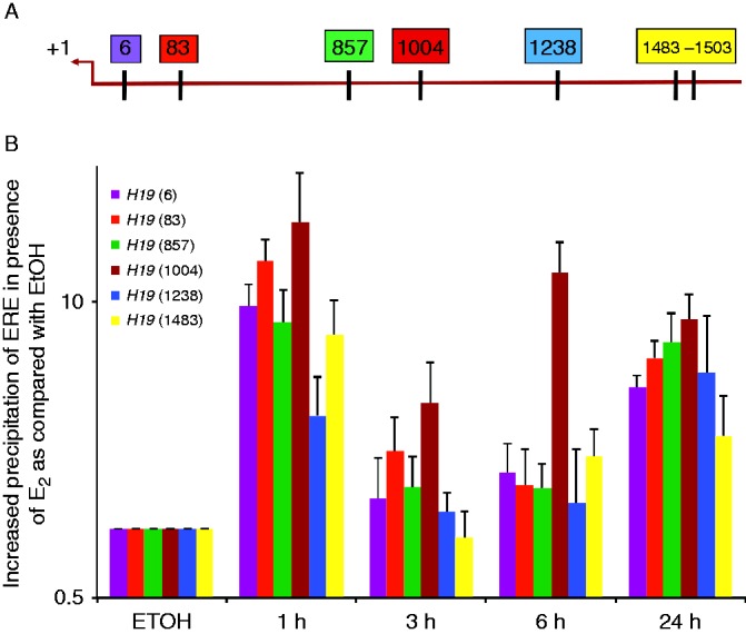
Figure 5.

ERα binds to H19 promoter through EREs. (A) This figure shows the location of EREs within 1500 base pairs of the transcription start site (TSS, +1) of the H19 proximal promoter. Each box is representative of one ERE half-site, and the number within each box shows its location away from the TSS. (B) Chromatin immunoprecipitation was performed to examine the ERα occupancy of each ERE. MCF7 cells were grown in estrogen-depleted media and treated either with EtOH or E2 for the indicated times. ERα bound to each ERE was quantified using qPCR. As shown, ERα binding to the H19 promoter was detectable after 1 h of exposure to E2.

 This work is licensed under a