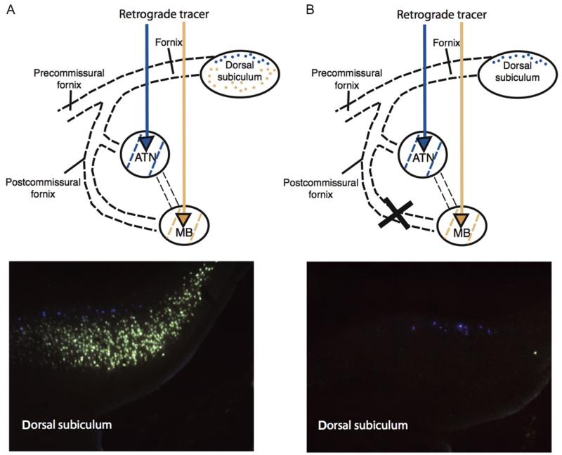
FIGURE 2.
Disconnection of the descending postcommissural fornix in rats. Rats underwent a control surgery (A) or a radiofrequency lesion of the descending postcommissural fornix (B). Fluorescent retrograde tracers were then injected into the mammillary bodies and anterior thalamic nuclei. In an intact rat (left), neurons containing the two retrograde tracers can clearly be seen. The deep subicular layers (Fast Blue (gray in the print version)) project to the anterior thalamic nuclei whereas the more superficial layers (Fluorogold (light gray in the print version)) project to the mammillary bodies. Following postcommissural fornix lesions, there is a complete disconnection of subicular-mammillary projections as shown by the loss of Fluorogold (light gray in the print version) label. This demonstrates that the hippocampal formation projects to the mammillary bodies solely by way of the fornix. Rats with these disconnection lesions are only very mildly impaired on spatial memory tasks (Vann, 2013; Vann et al., 2011).

