
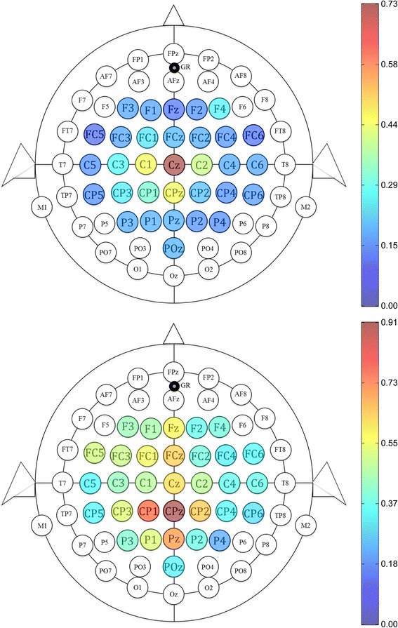
Fig. 7.
Importance of each EEG channel for dorsiflexion. Channels are colored based on their μ value, from dark blue (unimportant) to dark red (highly important). These two representative maps are taken from subject S8 (top) and S9 (bottom)
Official websites use .gov
A
.gov website belongs to an official
government organization in the United States.
Secure .gov websites use HTTPS
A lock (
) or https:// means you've safely
connected to the .gov website. Share sensitive
information only on official, secure websites.
Importance of each EEG channel for dorsiflexion. Channels are colored based on their μ value, from dark blue (unimportant) to dark red (highly important). These two representative maps are taken from subject S8 (top) and S9 (bottom)