
Fig. 3.
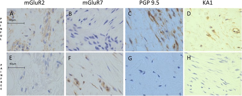
There were glutaminergic differences between painful and pain-free rotator cuff tendons. The expression of several glutaminergic markers was different between groups; mGluR2 and KA1 were increased in the painful group, while mGluR7 was increased in the pain-free group. mGluR7 staining was exclusively of endothelial cells, while mGluR2 and KA1 staining was of both endothelial cells and resident tendon cells. PGP 9.5 expression (type B synoviocyte marker) was increased in the painful group. Photomicrographs depict the immunohistochemical staining of mGluR2 (a, e), mGluR7 (b, f), PGP 9.5 (c, g) and KA1 (d, h). Scale bars: 40 μm. KA1 kainate receptor 1, mGluR metabotropic glutamate receptor, PGP9.5 protein gene product 9.5