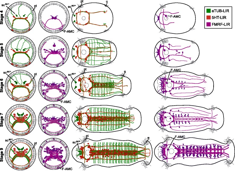
Fig. 11.

Diagrams of 5HT-LIR, aTUB-LIR and FMRF-LIR in C. teleta larvae. aTUB-LIR is shown in green, 5HT-LIR in red and FMRF-LIR in purple. The stomatogastric nervous system is omitted for clarity. The left two columns are anterior views with ventral down. The right two columns are ventral views with anterior to the left. Stage is indicated to the left of each row. F-AMC, FMRF-LIR anterior mouth cell; pt, prototroch; scac+, acetylated tubulin-positive sensory cells; tt, telotroch
