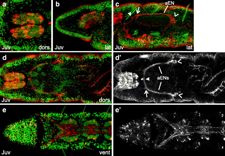
Fig. 9.

FMRF-LIR in 7-day old C. teleta juveniles. Images are z-stack confocal images of 7-day old juveniles labeled with anti-FMRF (red) and Hoechst nuclear stain (green). Panels labeled with an apostrophe (e.g., a') are single-channel images of FMRF-LIR from the merged image without an apostrophe (e.g., a). All panels are to the same scale unless otherwise noted. a is a cropped, 1.25x magnified image of the brain. b is a cropped view of the brain; c is a cropped view of the pharynx; d is a cropped view of the brain, pharynx and anterior portion of the midgut; e is a cropped view of the head and first two ganglia in the ventral nerve cord. In c and d’, open arrowheads point to clusters of neurons with FMRF-LIR near the pharynx (stomatogastric ganglia), a closed arrowhead points to a single cell with FMRF-LIR that is anterior to the medial portion of the pharynx, the open arrow indicates the first branchpoint in the anterior enteric nerve (aEN). Stage is indicated in the lower-left corner, and view is indicated in the lower-right corner. All lateral views are of the left side. Anterior is to the left in all ventral, dorsal and lateral views, and ventral is down in all anterior views. ant, anterior; dors, dorsal; lat, lateral; vent, ventral
