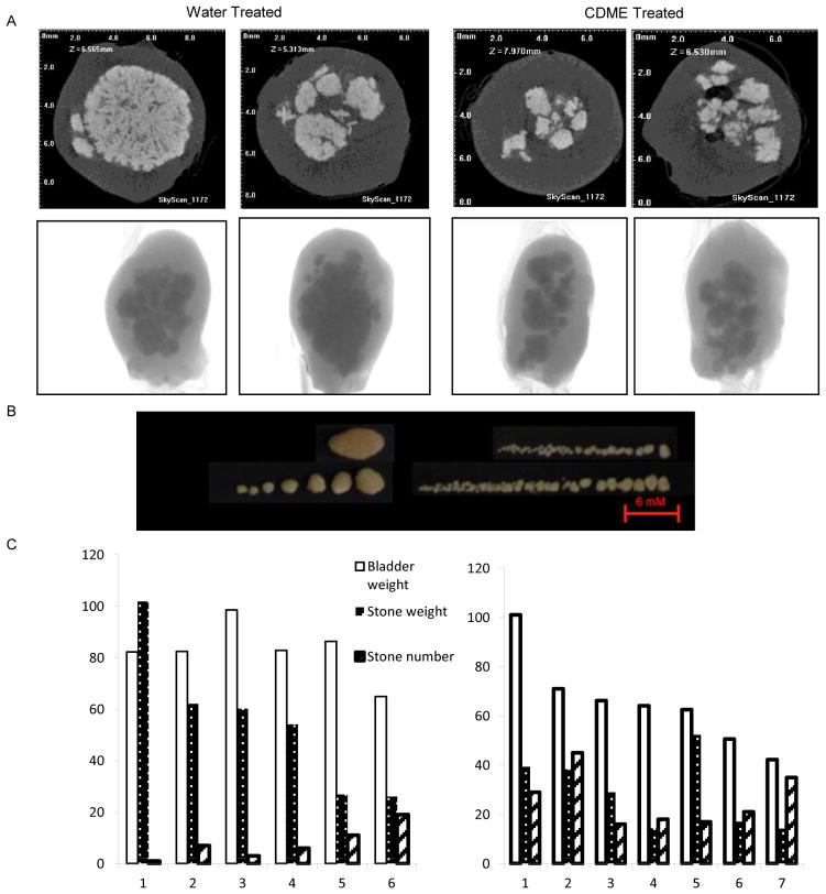
Figure 2. Analysis of cystine stones.
(A). Micro CT images of bladders from two Slc3a1 knockout mice treated with water (left panel) or CDME (right panel). For each mouse, the top panel is a cross-sectional image and the bottom panel is the intact organ. (B). Stones retrieved from the bladder of a water- or CDME-treated mouse. (C). Quantification of bladder weight, stone weight, and stone number in the water- and CDME-treated groups.

