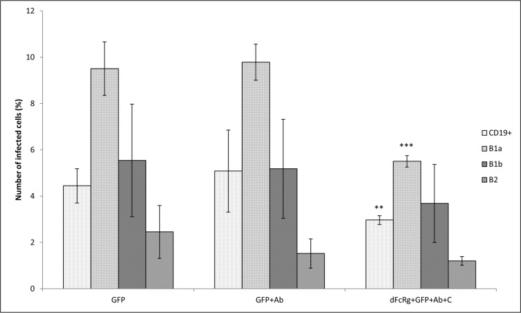
Fig 7. Blocking of FcγR.
Peritoneal cells were incubated with the antibody against CD16/32 (dFcRg). Thereafter, cells were infected with F. tularensis LVS/GFP (GFP), F. tularensis LVS/GFP opsonized with antibodies (GFP+Ab), and F. tularensis LVS/GFP opsonized with murine fresh serum and antibodies (dFcRg+GFP+Ab+C) at MOI 500. Entry into all B cells (CD19+) and individual B cell subsets was detected 3 h after infection by flow cytometry. Error bars indicate SD around the means of samples processed in triplicate. Two-tailed t-test was used to test for significant differences between GFP and GFP+Ab and between GFP+Ab and dFcRg+GFP+Ab+C (*** P < 0.001, ** P < 0.01). Results shown from one experiment are representative of three independent experiments

