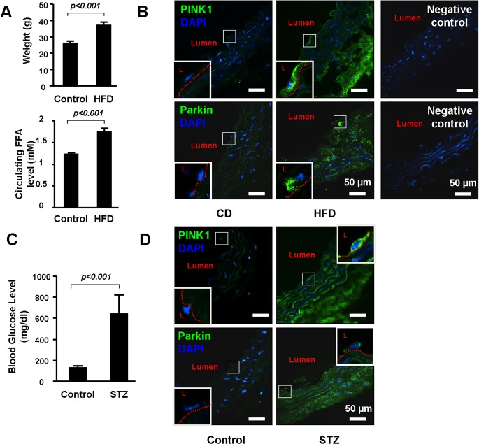
Fig 6. PINK1 and Parkin were up-regulated in the vascular wall of the obese and diabetic mice.
(A). Four week old wild-type male WT mice were either fed with chow diet (CD, n = 6) or high fat diet (HFD, n = 12) for 12 weeks. Body weight levels and free fatty acid (FFA) were compared between the groups. (B). Representative immunostaining showed increased PINK1 and Parkin in aortic wall and in endothelial cells (enlarged in the square box) in HFD fed mice. L indicated lumen. Red dot line indicated internal elastic plate of aorta. Negative controls for the PINK1 and Parkin immunostaining were shown on the right. (C). Eight week old wild-type male mice received daily IP injection of 50 mg/kg body weight streptozotocin (STZ, n = 8) or vehicle (n = 5) for five consecutive days. Aortas were collected 4 weeks after the STZ injection. Blood glucose level was compared between the groups. (D) Representative immunostaining showed increased PINK1 and Parkin in the aortic wall and in endothelial cells (enlarged in the square box) in STZ treated mice. L indicated lumen. Red dot line indicated internal elastic plate of aorta. Negative controls for the PINK1 and Parkin immunostaining were the same of (B).

