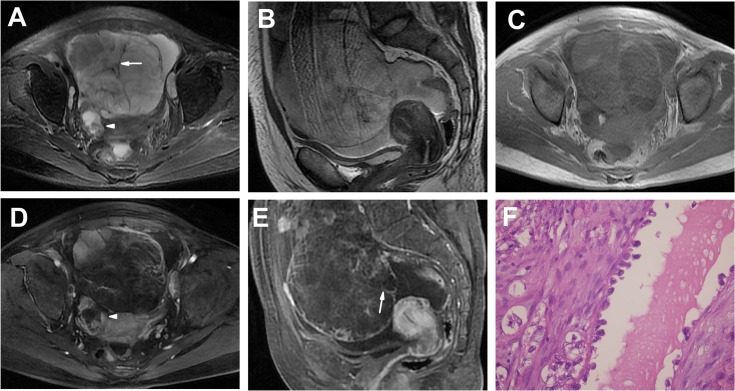
Fig 2. Bilateral OCCCs in a 47-year-old woman (Patient 10 in Tables 1 and 2) presenting with abdominal distension for 2 months.
A-C Axial (A), sagittal T2WI (B) and plain (non-contrast) T1WI (C) showing a left-side, large, well-defined multilocular cystic mass with a few irregular lumen solid protrusions and many irregular septations (> 3 mm, arrow) and similar MRI findings of a right-side, small, multilocular cystic mass with protrusions (arrow head). The SI of the cyst was high on T2WIs and low on T1WI. The solid protrusions had heterogeneous iso-slightly high SI on T2WIs and iso-SI on T1WI. Bulk ascites were detected. D, E Enhanced T1WIs showing markedly heterogeneous and prolonged enhancement solid protrusions and septations (arrow). F The tumor shows a cyst lined with hobnail cells. Secretions were found in the lumen of the cyst. (HE 40 & 10).

