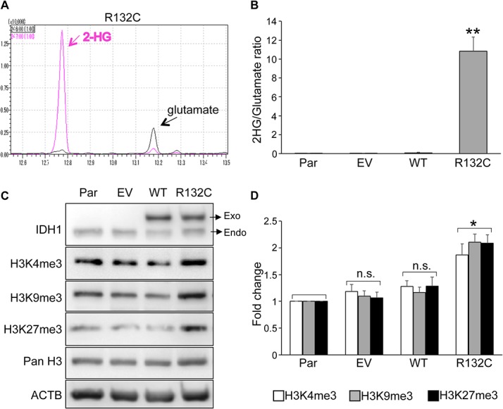
Fig 1. IDH1 R132C produced 2-HG and increased global histone methylation in hMSCs.

A. Detection of 2-HG by GC-MS. Intracellular 2-HG was detected in the extracts of BM01 cells expressing the IDH1 R132C gene. Peaks with m/z 246 and 247 were selected as quantification ions for glutamate and 2-HG, respectively. B. Relative amount of 2-HG in hMSCs. The amount 2-HG and glutamate was measured by GC-MS in cell extracts from each type of cell and the ratio was demonstrated. The mean ± SE from the results of three donors was shown. C. Expression of active (H3K4me3) and repressive (H3K9me3 and H3K27me3) histone marks. Protein lysates were prepared from each type of BM01 cell and used for western blotting to detect exogenous (Exo) and endogenous (Endo) IDH1 and indicated histone H3. D. Quantitative analyses of active and repressive histone marks. The expression level of each mark in infected hMSCs (EV, WT, or R132C) was demonstrated as a value relative to those in parental hMSCs (Par). The mean ± SE from the results of three donors was shown. Par, parental hMSC; EV, hMSCs infected with the empty vector; WT and R132C, hMSCs infected with the vector containing the wild-type IDH1 or IDH1 R132C gene, respectively. *, p<0.05 and **, p<0.01 by Dunnett`s multiple comparisons test compared to the parental cells.
