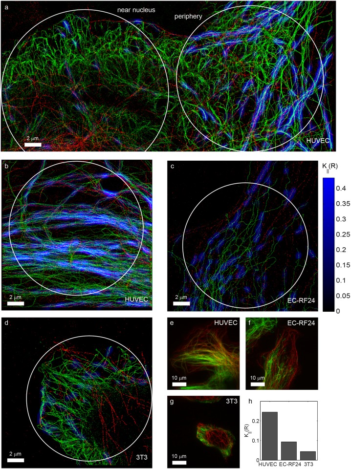
Fig 7. Co-orientation strength in endothelial and fibroblast cells.
Localization microscopy images of tubulin (red) and vimentin (green) in various cell types. (a) Large SR image of a HUVEC cell, showing that co-orientation is predominantly observed in the peripheral parts (right), and not near the nucleus (left). (b-d) Higher magnifications of comparable peripheral parts of (b) a HUVEC cell showing extensive co-orientation, (c) a EC-RF24 endothelial cell with less, but still significant co-orientation, and (d) a NIH-3T3 fibroblast as an example of a cell-type with very little co-orientation. (e-g) TIRF images corresponding to b, c and d and (h) quantification of the co-orientation strength for the circular ROIs in these three examples for R = 200 nm.

