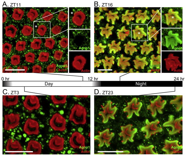
Figure 1. Light-mediated control of Agop1 cellular localization.
A. An Anopheles retina at ZT11, an hour prior to the initiation of the dark cycle, was probed for Agop1 (green) and actin (red). The location of the R7 photoreceptor cell body, lacking Agop1 expression, is marked by an asterisk in the highlighted ommatidial unit. Scale bars in all images are 20 μm.
B.Anopheles retina at ZT16, four hours after initiation of the dark cycle. After the light-dark transition, Agop1 (green) moves from the cytoplasmic region of photoreceptors surrounding the actin-rich rhabdom (red) to within the rhabdom. Agop1 is expressed in all peripheral cells (R1-6). Agop1 expression in the R8 photoreceptor is also evident in some central R8 rhabdomeres (asterisk in highlighted ommatidial unit at right).
C, D. Prior to dawn (ZT23), Agop1 rhodopsin localization remains within the peripheral rhabdoms (D), and moves completely to the cytoplasm by ZT3 (C), three hours after initiation of the light cycle.

