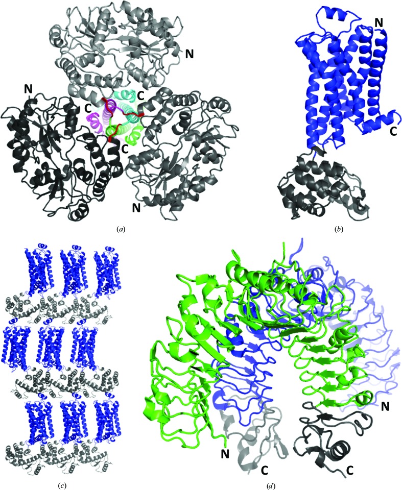
Figure 2.
Examples of successful application of the heterologous fusion-protein approach. The structures are not shown on the same scale. (a) Cartoon diagram of the structure of HTLV-1 gp21 (subunits are shown in different colours) fused at the N-terminus to MBP (in different shades of grey) with a three-Ala linker (red; PDB entry 1mg1; Kobe et al., 1999 ▸). (b) Cartoon diagram of the structure of β2-adrenergic receptor (β2AR; blue) with T4 lysozyme (T4L; grey) inserted into a loop in β2AR (Rosenbaum et al., 2007 ▸; PDB entry 2rh1). (c) A view of crystal-packing interactions for the β2AR-T4L fusion protein [shown and coloured as in (b)]. Note the crystal contacts between the fusion partner T4L and the soluble portion of β2AR. (d) Cartoon diagram of the structure of the complex of the extracellular domains of TLR1 (green) and TLR2 (blue) (Jin et al., 2007 ▸; PDB entry 2z7x). Both proteins are fused at the C-terminus to VLR as the fusion partner (grey). All structure figures were produced with PyMOL (Schrödinger).

