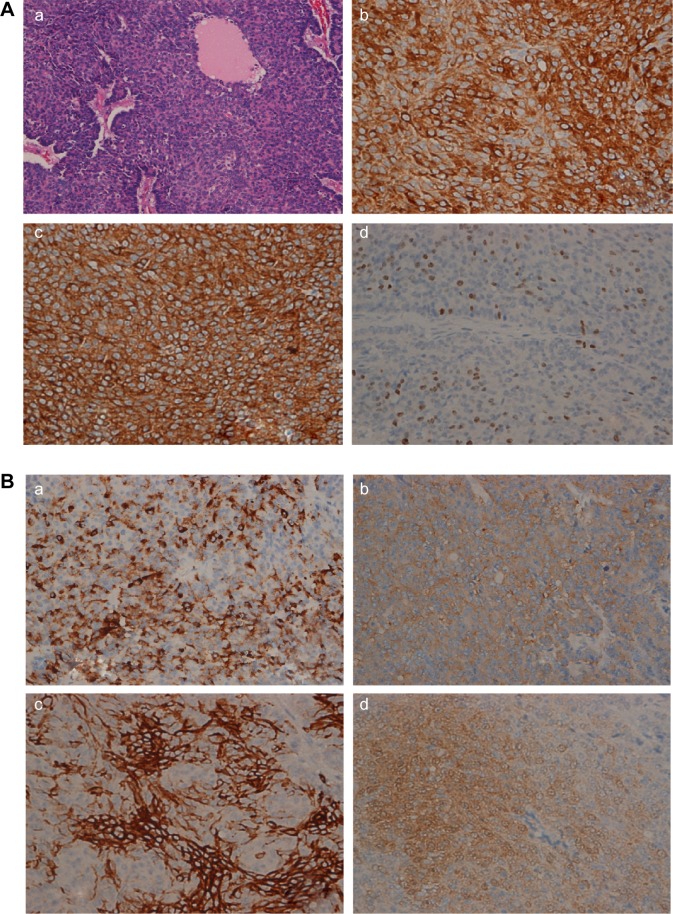
Figure 2.
Histopathology of the pancreas tumor.
Notes: (A) (a) Hematoxylin–eosin image of the pancreas tumor (HEX200). (b) Immunohistochemistry of trypsin (×400, Abcam). (c) Immunohistochemistry of chymotrypsin (×400, Abcam). (d) Immunohistochemistry of Ki-67 (×400, Abcam [Cambridge, MA, USA]). (B) (a) Immunohistochemistry of chromogranin A(×400, Abcam) (b) Immunohistochemistry of synaptophysin (×400, Abcam). (c) Immunohistochemistry of CD56 (×400, Abcam). (d) Immunohistochemistry of neuron-specific enolase (NSE) (×400, Abcam).

