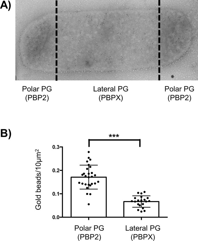
Fig 2. GM5 detection on polar and lateral PG.

A) Transmission electronic microscopy image of immuno-gold detection of GM5 using vancomycin labelling of N. bacilliformis saculli. B) Estimation of the mean numbers, with standard deviation, of gold beads by 10μm2 of sacculi surface of both polar/septal and lateral PG measured on around 20 cells (*** p≤0.001).
