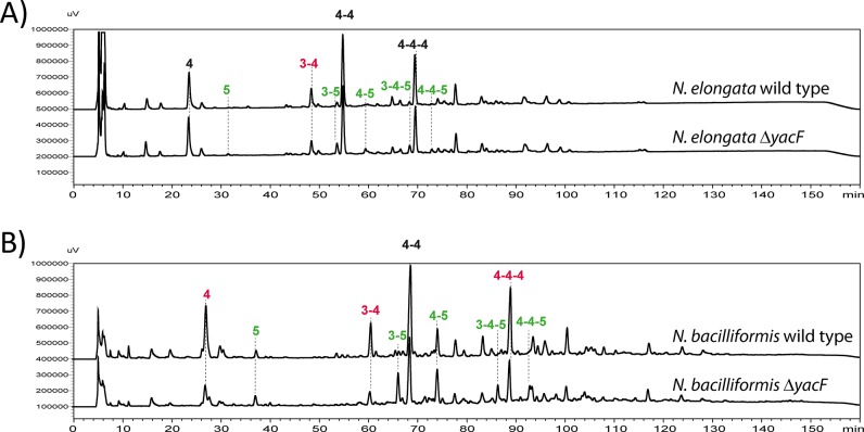
Fig 5. Increased proportion of pentapeptide muropetides in absence of YacF.
Reverse-phase HPLC analyses of muropeptides after mutanolysin digestion of purified insoluble PG of: A) N. elongata wild type and ΔyacF and B) of N. bacilliformis wild type and ΔyacF. The numbers indicate the different muropeptides; for example, 3 stand for GM3. The peak labeled 4–4 corresponds to a dimer of GM4 crosslinked by the two stem peptides. The colors indicate a, more than two fold, increase (green) or decrease (red) of the indicated muropeptide in the ΔyacF PG. This is representative chromatogram of experiments done at least three times. The raw quantification can be found in S3 Fig for N. elongata and the corresponding mutants.

