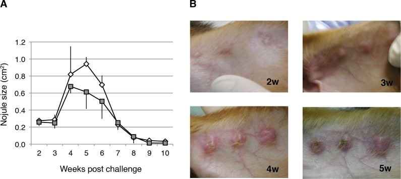
Fig 3. Time course of skin lesion development in dogs infected with L. major.
(A) Beagle dogs were infected intradermally (in the ears) with 5 × 10⁷ infective promastigotes of L. major per spot, and the lesion sizes were measured weekly. Parasite growth was evaluated as nodule size. Three independent spots per dog were determined and followed-up. Data are shown as means ± SEM, and the error bars reflect the three inoculated spots. (B) Images of lesions at 2 to 5 weeks after infection.

