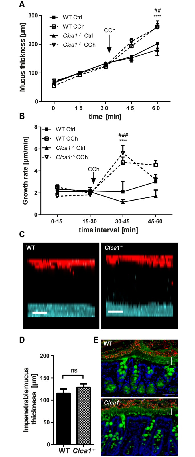
Fig 3. Clca1 -/- has no effect on mucus growth, responsiveness and penetrability in Clca1 -/- mice.

(A) After flushing distal colon explants, mucus thickness and growth were similar in Clca1 -/- mice (dotted lines) compared to WT (filled lines). After CCh stimulation, an increase in mucus thickness compared to unstimulated explants was observed both in WT and in Clca1 -/- mice. Ctrl = control. (B) The growth rate was constant during unstimulated conditions whereas a significant growth rate increase in response to CCh was evident in both groups shortly after its addition. No significant difference was observed between the groups. Ctrl = control. (C) Ex vivo mucus penetrability assessment using bacteria-sized beads (1 μm, red) and confocal microscopy was performed. Representative z-stack projections from WT and Clca1 -/- mucus 30 minutes after tissue mounting both showed a clear separation between the tissue (blue) and the beads (red). Scale bars 50 μm. (D) The impenetrable mucus thickness, measured as the distance between the tissue and the sedimented beads in the confocal z-stacks did not differ between WT and Clca1 -/- mice. ns = non-significant. (E) FISH with a general bacterial 16S probe (EUB338, red), counterstained for Muc2 (anti-MUC2-C3, green) and DNA (Hoechst 34580, blue) in sections from distal colon confirmed the impenetrability of the inner mucus layer both in WT and Clca1 -/- mice with a clear separation of the tissue and bacteria. i = inner mucus layer. Scale bars 100 μm. n = 5 per group. Data are presented as mean ± SEM. ## p < 0.01, ### p < 0.001 for WT Ctrl vs. WT CCh; ****p < 0.0001 for Clca1 -/- Ctrl vs. Clca1 -/- CCh.
