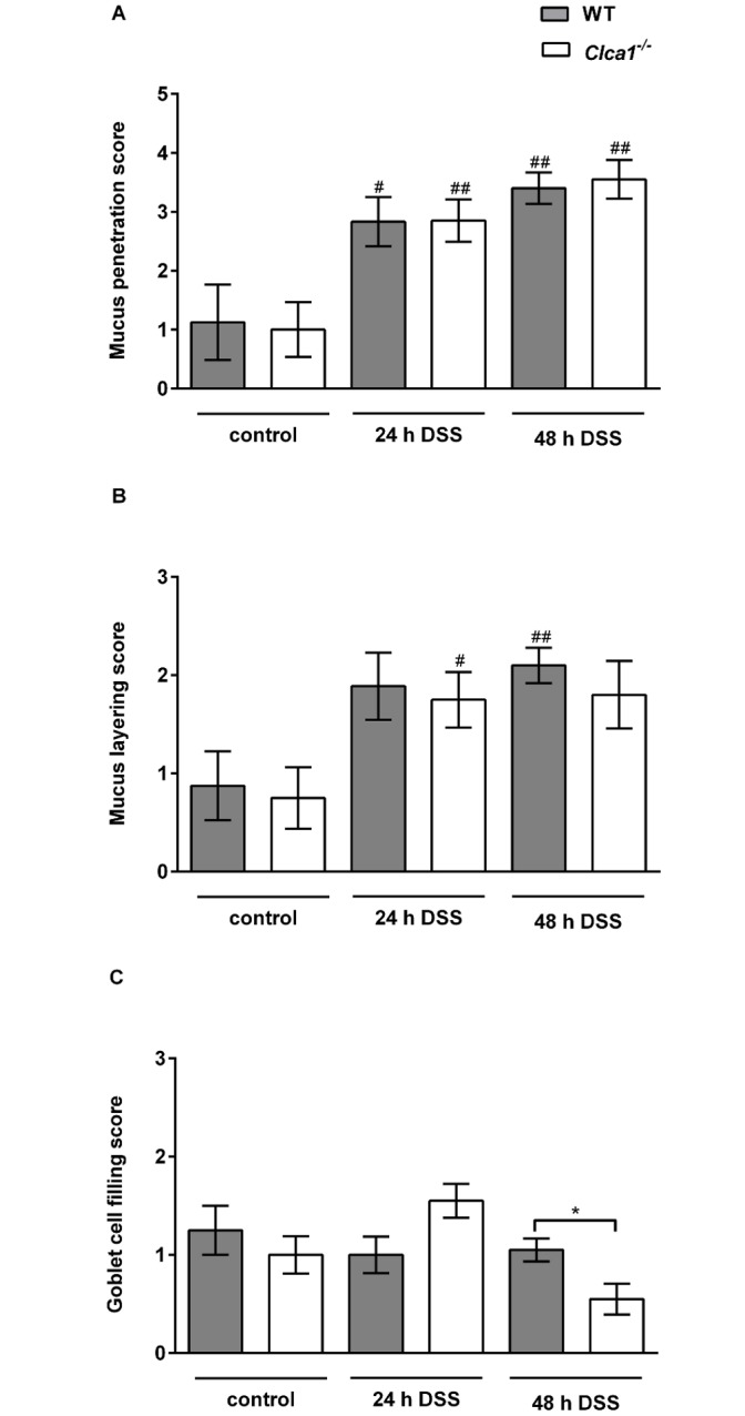
Fig 4. The mucus barrier of WT and Clca1 -/- mice are identically affected during DSS challenge.

Mucus penetration, mucus layering and goblet cell filling in the colon of mice after 24 and 48 h DSS treatment were assessed using IF microscopy. (A) Mucus penetration increased identically under DSS treatment, however, without any significant difference between the genotypes. (B) The mucus layering score also increased under DSS influence without any observable difference between the genotypes. (C) The goblet cell filling score did not show any statistically significant differences, neither between genotypes nor between naive vs. DSS-treated animals, except for WT vs. Clca1 -/- mice after 48 hours. Mean values (n = 8 to 10). The scoring system is depicted in S3 Table and S4 Table. #p < 0.05 and ##p < 0.01 versus the naive control group. *p < 0.05 as indicated. Scale bars 50 μm.
