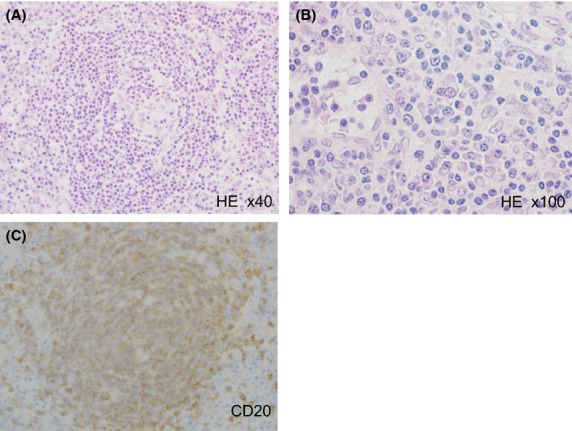
Figure 1.

Histological and immunohistochemical findings of the biopsied specimen from the right inguinal lymph node. Hematoxylin and Eosin (HE) staining showed that the biopsied lymph node comprised slightly-indistinct large hyperplastic follicles with expanded mantle zones. Endothelial hyperplasia was also observed in the follicle (A). Prominent plasma cell infiltration was also noted in the interfollicular areas (B). CD20 immunostaining indicated that the follicle comprised B lymphocytes (C).
