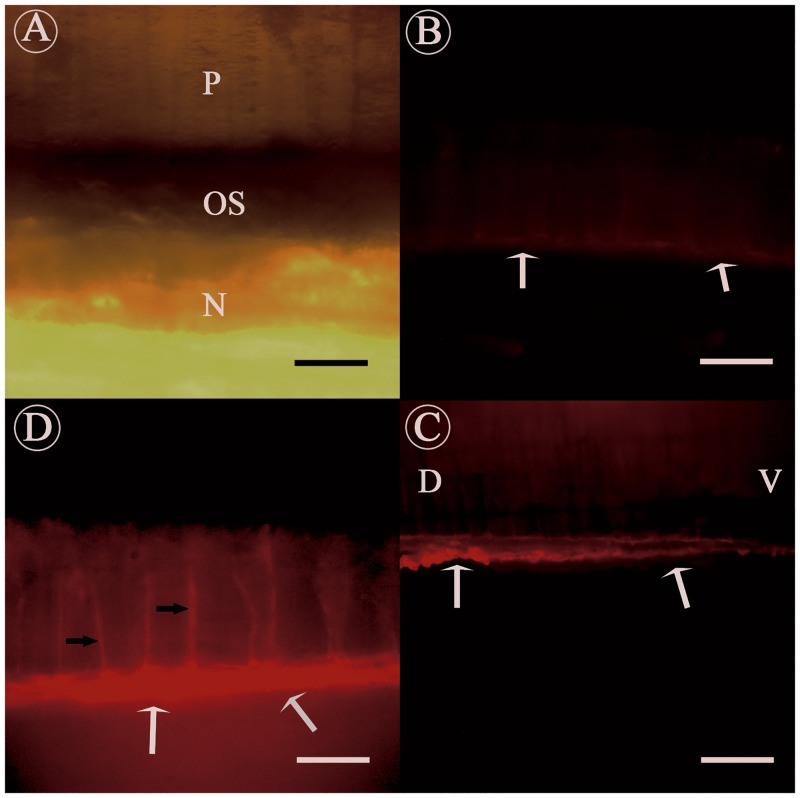
Fig 3. Immunofluorescence localization of native KRMP-3 in the adult shell of P. fucata, with anti-rKRMP-3 antibody.
Cleaned and modified shell was decalcified and incubated using anti-rKRMP-3 antibody (C, D) or preimmune serum (B). A, Light micrograph of the shell. The shell presents a representative nacroprismatic microstructure, with columnar calcitic prisms in the upper and nacreous layer in the lower. The two calcified layers are generally separated by an organic layer. P, prismatic layer; N, nacreous layer; OS, organic sheet. B, negative control staining with preimmune serum. The white arrows indicated only a small amount of background staining was detected in the organic sheet. C, staining with anti-rKRMP-3 antibody. The white arrows showed positive signal in the organic sheet. Meanwhile, the positive signal from the ventral to dorsal reflects the distribution of organic sheet; D, dorsal; V, ventral. D, high magnification of the sample. Positive signal detected not only in the organic sheet (white arrows), but also in the prismatic sheath (black arrows). Scale bars in (A, D), 12.5 μm and in (B, C), 50 μm.

