
Fig 5. Surface presentation of SRID mutations on HDAC4c structure.
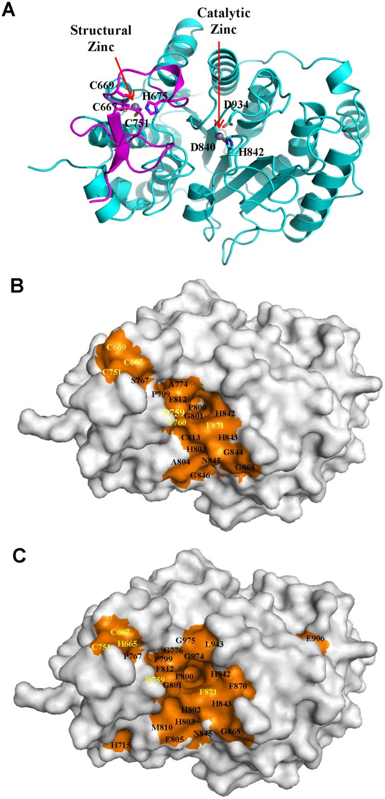
(A) Ribbon diagram of HDAC4c structure (PDB code 2VQJ) [40]. The structural zinc-binding domain is shown by magenta. Two zinc ions and their chelating residues are drawn as spheres and sticks, respectively. (B) Positions of SRID mutations of HDAC4c are presented on the surface of the HDAC4c structure (PDB code 2VQJ). Among SRID mutants of HDAC4c, 21 residues located on the surface are indicated, and their surface positions are shown in orange. (C) Surface presentation of SRID mutations of HDAC5c at the corresponding positions of the HDAC4c structure. The positions of the SRID mutations of HDAC5c were changed to those of HDAC4c according to MSA data, and presented on the surface of HDAC4c structure in orange. The residues specific to class IIa HDACs are indicated in yellow color.