Figure 2.


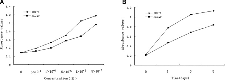
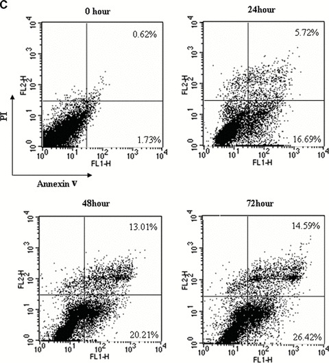
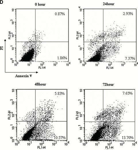
Effects of acitretin on apoptosis in SCL-1 cells. (A) SCL-1 cells and HaCaT cells were treated with acitretin at five different concentrations for 3 days. (B) SCL-1 cells and HaCaT cells were treated with 10−5 M of acitretin for various times. To determine and quantify the induction of apoptosis by acitretin in the SCL-1 cells and HaCaT cells, 20 μl of cell lysate was used for Cell Death Detection ELISA kit. DNA fragmentation was quantified at 405 nm. Data are shown as median of three different independent experiments. (C and D) SCL-1 cells (C) and HaCaT cells (D) were cultured with 10−5 M acitretin for 0, 24, 48 and 72 hrs. The cells were then stained with annexinV-FITC and PI labelling and were analysed by flow cytometry. The percentage of cells in each window is indicated. The percentage of annexinV-positive apoptotic cells in SCL-1 cells and in HaCaT cells both dramatically increased in a time-dependent manner. Three independent experiments were performed with similar results.
