
Figure 4.
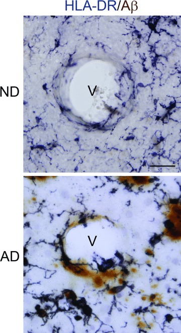
Representative double immunohistochemical staining for microglia (HLA-DR marker) and Aβ (6F/3D marker) in the entorhinal cortex of non-demented (ND) and Alzheimer’s disease (AD) tissue. Sections were incubated with HLA-DR and Aβ followed by visualization using DAB/nickel ammonium sulphate (anti-HLA-DR, dark purple colour) or DAB (anti-Aβ, brown colour). Staining shows blood vessels (v) with AD tissue demonstrating close association between Aβ, microglia and vessel. Scale bar represents 50 μm. Note the predominant ramified and ameboid morphologies of microglia in ND and AD tissue, respectively.