
Figure 6.
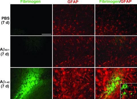
Fibrinogen and astrocyte (GFAP) double immunofluorescent staining in Aβ1-42-injected (7 days) hippocampus. GFAP immunoreactivity (ir; middle panels) was increased with Aβ1-42 compared to controls (PBS and Aβ42-1). Typical merged staining is shown in the right panels. Scale bar = 200 μm. The inset in the lower right panel presents a higher magnification showing negligible association of fibrinogen with GFAP ir astrocytes.