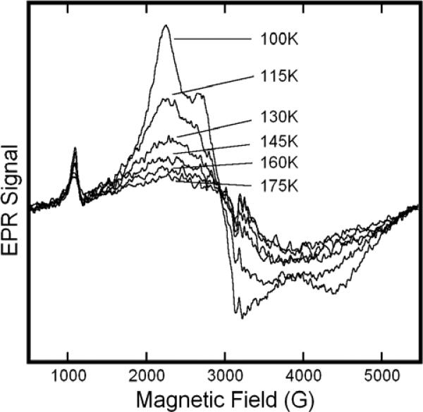
Figure 5.
Variable Temperature EPR spectra of solutions of metHb:NO2− in HEPES buffer at pH 7.4. The sample included metHb at 0.65M and a twenty-fold molar excess (per heme) of NaNO2−. Spectra were obtained at the indicated temperatures in the range 100K to 175K. EPR spectra were recorded at 9.1 GHz with a 16.67 G/s sweep rate, a 0.5 s time constant, 5 G modulation amplitude, and 10 mW of microwave power.

