
Figure 4. TGF-β1 via Smad3, and AhR support the conversion of TH17 to TR1.
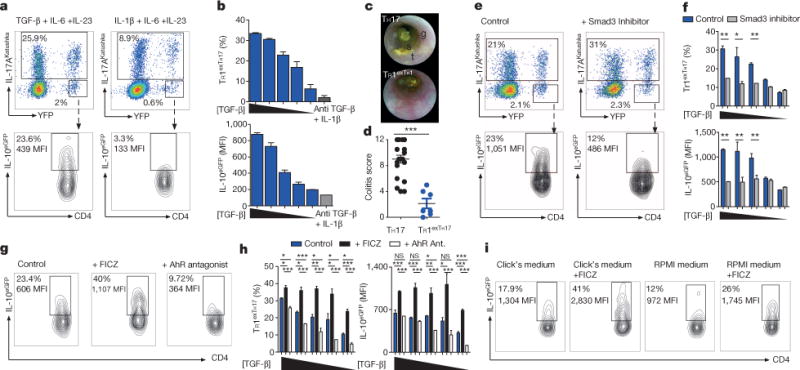
TH17 cells were differentiated in vitro in the presence of IL-6, IL-23 with TGF-β1 or with IL-1β and anti-TGF-β monoclonal antibody. TGF-β1 was diluted 1:2 starting from 4 ng ml−1. a, Flow cytometric analysis of TH17, exTH17 and TR1exTH17 (gated on exTH17). b, Percentages and IL-10 MFI of TR1exTH17 cells. Technical replicates (n = 2) of one experiment out of seven. c, d, Endoscopic pictures and score of mice injected with TH17 or TR1exTH17 cells polarized with TGF-β1. Stool inconsistency (s), increased mucosal granularity (g) and a lack of translucency (t). Each dot denotes one biological replicate. Mean and s.e.m., ***P ≤ 0.0005 by Mann Whitney U-test, two tailed. e, Flow cytometric analysis of TH17, exTH17 and TR1exTH17 cells cultured in the presence of TGF-β1 (diluted as above), IL-6, IL-23 ± Smad3 inhibitor. f, Percentages and IL-10 MFI of TR1exTH17 cells. Technical replicates (n = 3) of one experiment out of five are shown. Mean and s.e.m., *P ≤ 0.05, **P ≤ 0.005 by paired t-test. g, Flow cytometric analysis of TR1exTH17 (gated on exTH17) cultured in the presence of TGF-β1 (diluted as above), IL-6, IL-23 ± AhR ligand (FICZ) or AhR antagonist. h, Percentages and IL-10 MFI of TR1exTH17 (gated on exTH17). Technical replicates (n = 3) of one experiment out of five are shown. Mean and s.e.m.; *P ≤ 0.005, **P ≤ 0.005, ***P ≤ 0.0005 by ANOVA (Tukey’s multiple comparison test). NS, non-significant. i, Flow cytometric analysis of TR1exTH17 cells cultured in the presence of TGF-β1, IL-6, IL-23 ± FICZ in the indicated medias. One experiment of two is shown.