Figure 1.
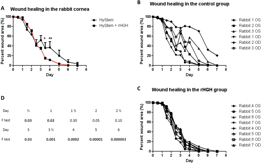
rHGH promotes corneal epithelial wound closure in vivo. A: Percent remaining epithelial defect (baseline being 100%) during 7 days in the control (HyStem alone) and rHGH (HyStem + rHGH) groups. Repeated measures ANOVA revealed p<.0001 for the interaction of time and rHGH treatment, and Bonferroni multiple comparisons showed significant difference between control and rHGH groups after 3 ½ and 4 days (*p<.05, **p<.01). B and C: percent wound area during 7 days in individual rabbit eyes in the HyStem and rHGH groups, respectively. D: F test values between control and rHGH groups at each individual time point, with values <.05 in boldface.

