Figure 3.
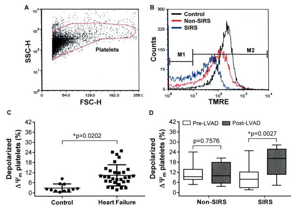
The change in % depolarized ΔΨm platelets among the Non-SIRS and SIRS groups before and after CF-LVAD implantation. (A) FSC versus SSC dot plot. The gate indicates position of platelets. (B) The distribution histograms show the change in TMRE fluorescence in the control, Non-SIRS and SIRS groups. (C) Scatter plot representing the differences in % depolarized ΔΨm platelets between the healthy controls and the HF patients at the baseline. *p<0.05 is considered significant in Student’s t-test. (D) Box-whisker plot shows the differences in % depolarized ΔΨm platelets before and after CF-LVAD implantation in the Non-SIRS and SIRS groups. The lines across each box plot represent the median value. The lines that extend from the top and the bottom of each box represent the lowest and highest observations still inside the lower and upper limit of confidence. *p<0.05 is considered significant in Mann–Whitney U-test.

