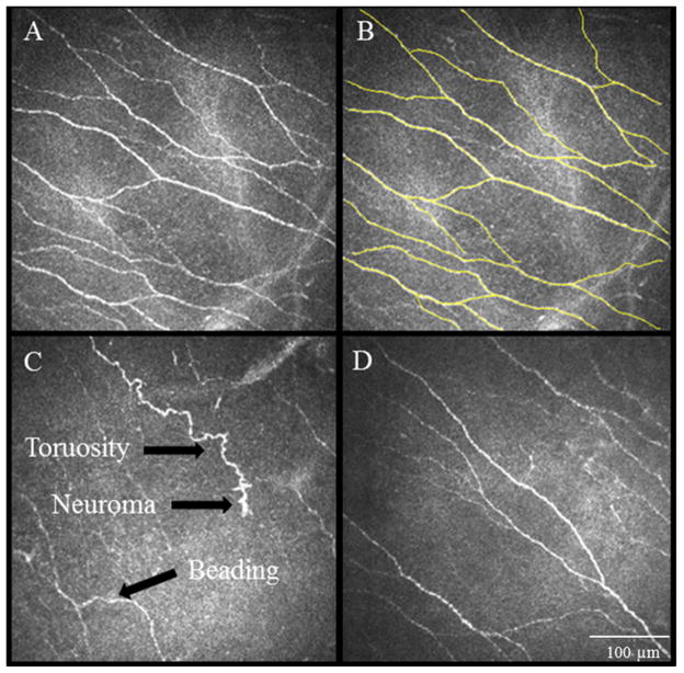
Figure 1.
IVCM images obtained at the level of the corneal subbasal nerve plexus demonstrate nerve alterations in patients of corneal neuropathy. A. Normal corneal subbasal nerve plexus. B. Nerve tracings using Neuron J to quantify the density of nerves. C. IVCM images showing significantly decreased corneal subbasal nerve plexus at baseline, prior to autologous serum tears therapy. Note the decrease in length and number of nerves, increased tortuosity, reflectivity, beading and formation of neuromas. D. IVCM image showing post-treatment findings of the patient shown at panel C. Note improvement in the plexus, still abnormal as compared to normal controls. Size bar=100μm.

