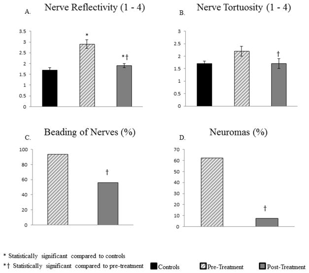
Figure 5.
Comparison of the subbasal nerve plexus morphology in patients suffering from corneal neuropathy, controls, and following treatment with ASTs. Bar graphs showing corneal nerve alterations in pre-treatment and post-treatment eyes in patients and in the control group. A. Reflectivity of nerves. B. Tortuosity of nerves. C. Beading of nerves. D. Neuromas. *P<.001, compared to the control group by analysis of variance, †P<.001, compared to the pre-treatment group by paired t-test.

