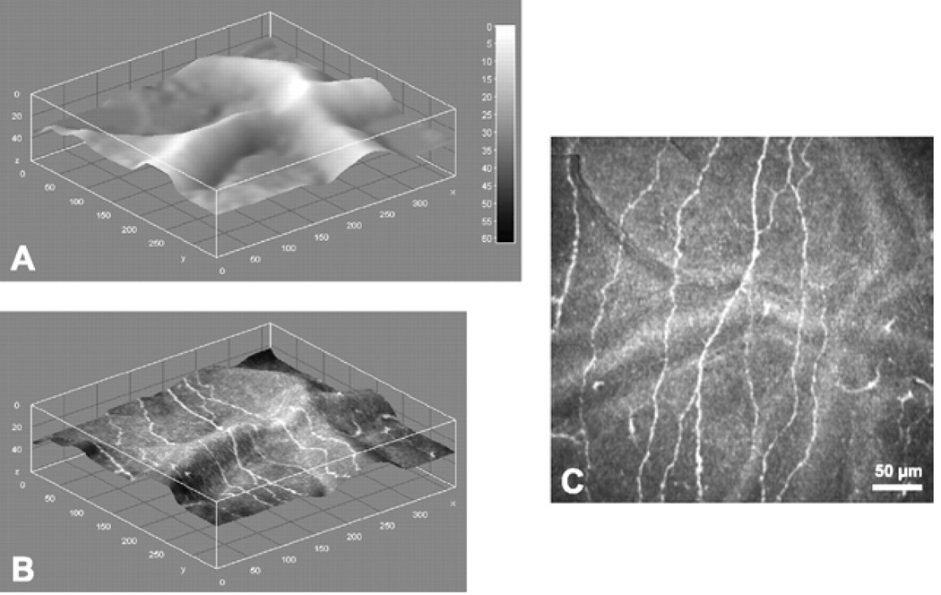
Figure 11.
Depth map and nerve fiber layer extraction at SNP level. A. Depth map of SNP layer inside the reconstructed volume. B. Depth map of SNP layer, textured with reconstructed image. C. Reconstructed image of the SNP. Image size: 362 × 347 pixels, 377 × 361 µm, ~85% of original image size. (Reprinted with permission from Allgeier S, Zhivov A, Eberle F, et al. Image reconstruction of the subbasal nerve plexus with in vivo confocal microscopy. Invest Ophthalmol Vis Sci 2011;52:5022-28.)

