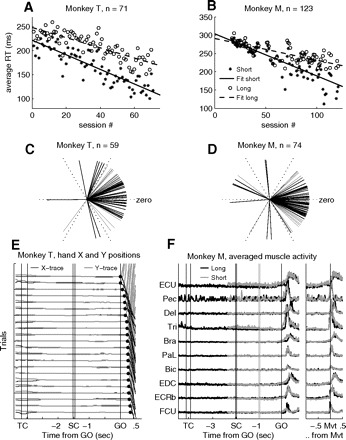
Fig. 2.

A and B: average RTs in short (stars) and long (open circles) delay trials in each recording session, plotted chronologically for monkey T (A) and monkey M (B). The lines are least-square fits to the data. C and D: difference between short and long trials (circular distance) of preferred directions (direction with the fastest RTs in average) for monkey T (C) and monkey M (D) for all recording sessions where all 6 movement directions were included; 57/59 and 59/74 of the sessions were significantly directionally selective in RTs in either short or long trials for monkeys T and M, respectively. The more the lines point toward zero, the smaller the circular difference in behavioral preference in short and long trials. E: example of hand X and Y positions for monkey T, from multiple correct repetitions of a long trial type. The x-axis indicates the timing of events. Each pair of black (x-direction of movement) and gray (y-direction of movement) lines corresponds to one trial (trials are displaced in steps of 0.5 cm on the y-axis for clarity). Filled circles after GO indicate movement onset. Trials are sorted off-line for increasing RT. F: averaged muscle activity from 10 arm and upper body muscles on the active side, recorded in representative and dedicated sessions in monkey M. Activity is aligned to the signals, at the left, and to movement onset (Mvt), at the right. Short trials are shown in gray, long trials in black, both for movements to the lower right (T2). The time of spatial cue (SC) onset and offset in long trials is indicated by 2 vertical black lines. The 2 vertical gray lines indicate SC in short trials. The selected muscles were (from top to bottom): extensor carpi ulnaris (ECU), pectoralis (Pec), deltoideus (Del), triceps brachii (Tri), brachioradialis (Bra), palmaris longus (PaL), biceps brachii (Bic), extensor digitalis communis (EDC), extsensor carpi radialis brevis (ECRb), and flexor carpi ulnaris (FCU). Artifacts attributed to the heartbeat can be observed on the electromyogram (EMG) taken from the pectoralis muscle.
