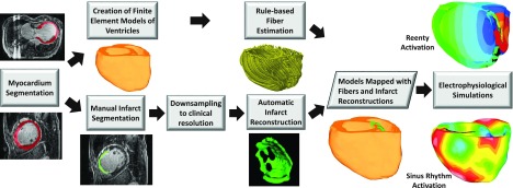
FIG. 2.

Block diagram for generation of models of individual hearts from LGE-CMR images for our electrophysiological simulation studies. Infarct reconstructions were performed as in Fig. 1. For each patient, two ventricular models, one with manually generated infarct reconstruction and the other with computed infarct reconstruction, were built. Ventricular fiber orientations were estimated using a rule-based method. The outcomes of electrophysiological simulations with computed infarct reconstructions were compared to those with manual reconstructions.
