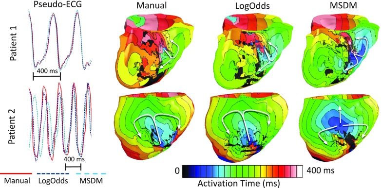
FIG. 6.
Comparison of pseudo-ECGs and activation maps for one beat of VT simulated for two patient hearts, with models that incorporate different infarct reconstructions. Activation maps are shown in septal and anterior views of the ventricles for Patients 1 and 2, respectively. The arrows highlight reentrant propagation patterns.

